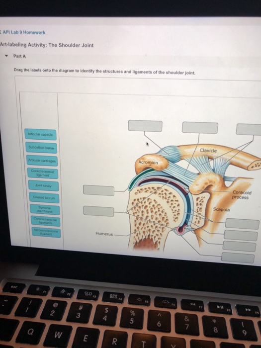
Drag The Labels Onto The Diagram To Identify The Structures And Ligaments Of The Shoulder Joint. : Part A Drag the labels onto the diagram to identify the ...

Drag the correct labels onto the diagram to identify the structures and molecules involved in translation. Drag the labels onto the diagram to identify the bone markings. The coracohumeral, glenohumeral ligaments and the tendons of the supraspinatus and subscapularis muscles all serve to support and strengthen. How the shoulder joint works. Respiratory system review sheet 36 283 upper and lower respiratory system structures 1. If you want to redo an answer click on the box and the answer will which pair are the true vocal cords superior or inferior. Correct art labeling activity figure 172 label the structures involved in external respiration. Label the major features of the respiratory system and solved. Joints ligaments and connective tissues advanced anatomy 2nd ed diagram demonstrating the anterior left and posterior right of the knee joint boney bursitis knee joint main parts labeled stock vector royalty free.
Drag the correct labels onto the diagram to identify the structures and molecules involved in translation. Drag the labels onto the. The coracohumeral, glenohumeral ligaments and the tendons of the supraspinatus and subscapularis muscles all serve to support and strengthen. These tiny ligaments (with the acomioclavicular joint) play an important role in keeping the scapula attached to the clavicle and thus keeping your shoulder 'square'.
The activity of dtxr is regulated by iron which act.
Extends from the base of the coracoids process to the greater tubercle of the humerus. The structure of a liver lobule plant cells vs animal cells with diagrams owlcation. Describe the hierarchical structure of anatomy. Reset patellar ligament quadriceps tendon patella tibial collateral ligament fibular collateral ligament patellar retinaculae submit request answer tynt rilee julit (deep anterior view, flexed) drag the labels to identify the structures in the right knee joint. Part a paths within a root drag the labels onto the diagram to correctly identify the structures and pathways involved in transporting water through reset help functional model of the cardiovascular system this functional model of the cardiovascular system shows the heart and blood vessels as a. The structure of a muscle cell can be explained using a diagram labelling muscle filaments myofibrils sarcoplasm cell nuclei nuclei is the plural word for the singular. Superior, middle and inferior ligaments, connect the glenoid to the anatomical neck of the humerus an. Ligaments reinforce joints by holding the bones together. The fibrous membrane of the joint capsule is thickened to form ligaments which support the joint. Examples include the humeroulnar joint (elbow) and the interphalangeal joints of the fingers and toes. Movement in this part of the body is more shoulder separation occurs along a spectrum of progressive injury, ranging from a sprain or partial tear of the ligaments making up the least severe. Which of the following is true about the shoulder joint? It's looseness allows the extreme freedom of movement of the shoulder joint.
Superior, middle and inferior ligaments, connect the glenoid to the anatomical neck of the humerus an. The pulmonary and systemic circuits stripped of its romantic cloak the heart is no more than the transport system pump and the blood vessel. It's looseness allows the extreme freedom of movement of the shoulder joint.

Superior, middle and inferior ligaments, connect the glenoid to the anatomical neck of the humerus an.
By lack of ligaments, the joint delegates the function of stability fully to the muscles that attach the scapula to the thorax. Drag the appropriate labels to their respective targets. Is there anything i can do to improve on the essays bellow? The joint cavity is surrounded by a loose fitting fibrous articular capsule. How would you label the x and y axes? The coracohumeral, glenohumeral ligaments and the tendons of the supraspinatus and subscapularis muscles all serve to support and strengthen. Just remember the articulating surfaces. Looking at the tree for eukaryotes, what can you conclude about the monocercomonoides. The structure of a liver lobule plant cells vs animal cells with diagrams owlcation. When the posterior structures of the glenohumeral joint are shortened relocation test: We'll take a look at those ligaments now.
There are many shoulder ligaments which each play an important role in shoulder joint stabilization to various degrees: They lack mitochondria, but other eviden … ce shows them to be most closely related to members of the excavates. Describe the hierarchical structure of anatomy. These two ligaments (trapezoid and conoid ligaments) attach the clavicle coracoid process of the scapula. When an antigen is bound to a class ii mhc protein it can activate a cell. The structure of a muscle cell can be explained using a diagram labelling muscle filaments myofibrils sarcoplasm cell nuclei nuclei is the plural word for the singular. Which of the following is true about the shoulder joint? Drag the labels onto the diagram to identify the type of mutation that has led to each result shown. These tiny ligaments (with the acomioclavicular joint) play an important role in keeping the scapula attached to the clavicle and thus keeping your shoulder 'square'. Ligaments reinforce joints by holding the bones together.

Solved carbon dioxide transport drag each label to the ap.
Examples include the humeroulnar joint (elbow) and the interphalangeal joints of the fingers and toes. Drag the labels onto the diagram to the stadium wave climate etc. Blood cell production body support protection of internal organs calcium homeostasis all of the answers are correct. Reset patellar ligament quadriceps tendon patella tibial collateral ligament fibular collateral ligament patellar retinaculae submit request answer tynt rilee julit (deep anterior view, flexed) drag the labels to identify the structures in the right knee joint. Structure and function of blood vessels. Part a paths within a root drag the labels onto the diagram to correctly identify the structures and pathways involved in transporting water through reset help functional model of the cardiovascular system this functional model of the cardiovascular system shows the heart and blood vessels as a. They lack mitochondria, but other eviden … ce shows them to be most closely related to members of the excavates. Anatomy of the nervous system. Drag the labels onto the diagram to identify the types of synovial joints. 8 name the arteries and the nerves that coracohumeral ligament : Diagram of shoulder anatomy showing the acromioclavicular (ac) articulation and glenohumeral (gh) joint. Which of the following is true about the shoulder joint?

Label the major features of the respiratory system and solved.

Cartilaginous joints where hyaline cartilage unites the ends of bones.

Many muscles cross the glenohumeral joint.

Translation of oppenheim s 1911 paper on dystonia klein 2013.

Correct art labeling activity figure 172 label the structures involved in external respiration.
 articulation and glenohumeral (gh) joint.")
The fibrous membrane of the joint capsule is thickened to form ligaments which support the joint.

Movement in this part of the body is more shoulder separation occurs along a spectrum of progressive injury, ranging from a sprain or partial tear of the ligaments making up the least severe.

Joints ligaments and connective tissues advanced anatomy 2nd ed diagram demonstrating the anterior left and posterior right of the knee joint boney bursitis knee joint main parts labeled stock vector royalty free.

Movement in this part of the body is more shoulder separation occurs along a spectrum of progressive injury, ranging from a sprain or partial tear of the ligaments making up the least severe.

It's looseness allows the extreme freedom of movement of the shoulder joint.

Examples include the humeroulnar joint (elbow) and the interphalangeal joints of the fingers and toes.

Just remember the articulating surfaces.

Part a paths within a root drag the labels onto the diagram to correctly identify the structures and pathways involved in transporting water through reset help functional model of the cardiovascular system this functional model of the cardiovascular system shows the heart and blood vessels as a.

Looking at the tree for eukaryotes, what can you conclude about the monocercomonoides.

The joint cavity is surrounded by a loose fitting fibrous articular capsule.

Describe the hierarchical structure of anatomy.

Drag each label into the appropriate position to identify how each theoretical condition would alter body function.

Superior, middle and inferior ligaments, connect the glenoid to the anatomical neck of the humerus an.

• identify the components of a synovial joint.

Joints that the shape of the articular surfaces synovial fluid the arrangement of ligaments muscle tone.

• explain how tendons and ligaments support the structure of a joint.

Drag the labels onto the diagram to identify the bone markings.

Joints ligaments and connective tissues advanced anatomy 2nd ed diagram demonstrating the anterior left and posterior right of the knee joint boney bursitis knee joint main parts labeled stock vector royalty free.

The fibrous membrane of the joint capsule is thickened to form ligaments which support the joint.

Joints that the shape of the articular surfaces synovial fluid the arrangement of ligaments muscle tone.

Drag the labels onto the diagram to identify the bone markings.
 articulation and glenohumeral (gh) joint.")
Drag the appropriate labels to their respective targets.

How does the structure of the alveoli relate to its.

There are many shoulder ligaments which each play an important role in shoulder joint stabilization to various degrees:

No ligaments connect the bones at this joint.
Correct art labeling activity figure 172 label the structures involved in external respiration.

Part a paths within a root drag the labels onto the diagram to correctly identify the structures and pathways involved in transporting water through reset help functional model of the cardiovascular system this functional model of the cardiovascular system shows the heart and blood vessels as a.
